by
Lauren Dubinsky, Senior Reporter | April 16, 2018
From the April 2018 issue of HealthCare Business News magazine
Making inroads into heart disease diagnosis
Two cardiac imaging technologies are in development that could open new avenues for predicting the risk of and diagnosing heart disease.
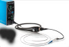
The EU’s Horizon 2020 collaboration is working on a handheld scanner that general practitioners can use during a routine health exam to spot patients with early-onset cardiovascular disease.


Ad Statistics
Times Displayed: 174457
Times Visited: 3181 For those who need to move fast and expand clinical capabilities -- and would love new equipment -- the uCT 550 Advance offers a new fully configured 80-slice CT in up to 2 weeks with routine maintenance and parts and Software Upgrades for Life™ included.
The device leverages a technique called Laser Doppler Vibrometry to create a vibration map of the chest and heart area. That map reveals signs of cardiovascular disease such as plaque build-up, arterial stiffness, arterial stenosis and heart dyssynchrony.
The current methods for diagnosing this condition are cardiac biomarkers, cardiac catheterization, chest X-ray, ECG, Holter monitoring and cardiac MR. Those approaches can be pricey, but the handheld scanner is expected to cost less than $2,000.
Medtronic has been appointed the technical coordinator of this collaboration and a prototype is expected to be unveiled this summer.
The University of Warwick in England, along with the Baker Heart and Diabetes Institute and Monash University in Australia, developed a laser imaging technique that can identify high-risk arterial plaques.
The team of researchers found that when they increased the wavelength of light that’s used to assess fatty build-up in arteries, they can identify rupture-prone deposits that often lead to blood clots, heart attack and stroke.
Current imaging techniques can identify some characteristics of high-risk plaques, but no method has been deemed reliable for selectively detecting these types of plaques. For instance, coronary angiography only detects narrow segments in the coronary artery.
If the laser imaging technique demonstrates positive results in clinical trials, it could one day be used to evaluate unstable fatty arterial plaques. It could also be used to monitor the effectiveness of drugs intended to prevent heart attack and stroke.
Back to HCB News