
Siemens Healthineers ACUSON
Bonsai cardiac ultrasound system.
Bonsai cardiac ultrasound system.
Imaging innovation yields new insights into cardiac health
April 16, 2018
by Lauren Dubinsky, Senior Reporter
Improvements in imaging technology are not only allowing cardiologists to evaluate patients in ways they never could before, through automation these new tools are simplifying the exam process and saving hospitals valuable time.
Guidelines from the American Society of Echocardiography call for 3-D chamber quantification on all patients undergoing an echocardiogram, but cardiologists have sometimes been known to overlook this time-consuming process when speed and efficiency are top priorities.
Conventional quantification involves the physician hand tracing to evaluate ventricular function, which typically takes about three minutes. There are also limitations associated with that method, such as foreshortening and not being able to visualize the entire endocardial border.
This is a prime example of an area where automated tools can simplify the imaging process and enable reproducible results. Meanwhile, the burden on the provider is significantly diminished.
“When you use automated intelligence, once you have acquired the data it doesn’t matter if a physician of two years or a physician with 20 years of experience presses the button because it will give you the same result,” says Dr. Roberto Lang, professor of medicine and director of the cardiac noninvasive imaging lab at the University of Chicago Medicine.
He uses Philips’ Anatomical Intelligence HeartModel tool, which is integrated into his EPIQ cardiac ultrasound systems. It performs 3-D cardiac chamber quantification by simultaneously computing the left ventricle and left atrium volumes from a single volume loop.
“When you have a volume of data, you have more images so you can see more pathology,” says Cassandra Gipson, clinical analyst at MD Buyline. “When you just have 2-D images, it’s limited because it’s a simple still image. With 3-D, you can turn the images around in any direction and see how deep the pathology is.”
Automated cardiac ultrasound imaging also improves workflow in the cardiac catheterization lab because it allows the technologist to have more time with the patient instead of having to stop and reformat.
Lang and his colleagues compared quantification between conventional 2-D exams and HeartModel and found that it reduced the time to obtain results by 82 percent, so if a cath lab typically treats 50 patients per day, he believes they could save over an hour with automation tools.
From a market standpoint, Philips is the leader in the cardiac ultrasound automation tool market and GE Healthcare follows closely behind, according to MD Buyline. Samsung Healthcare, Canon Medical Systems Corporation and Hitachi Healthcare Americas also offer this software on their premium cardiac ultrasound systems.
Ramping up investment and manufacturing of these automated, intelligent solutions is inevitable, according to Dr. Albert Hsiao, an assistant professor of radiology at the University of California San Diego.
“The sheer amount of information available to us through medical imaging has been on a steep incline, while the number of physicians available to handle this work is essentially flat,” he says.
A lower price point for advanced cardiac imaging
In March 2017, at the American College of Cardiology Annual Scientific Session and Expo, Siemens Healthineers unveiled a portable cardiac ultrasound system with high-level capabilities. The laptop-based ACUSON Bonsai is equipped with several of the features found on the company’s SC2000 premium cardiac ultrasound platform.
“This is to meet the needs of the routine echo space,” says Marti McCulloch, cardiology segment director for North America for Siemens Ultrasound. “When you look at the data, there are 34 million echos done per year. A lot of hospitals are moving toward more portable studies at the bedside.”
Also, when hospital administrators look at staff reductions as a way to curb spending, McCulloch says the transporters are usually the first to go. If a hospital doesn’t have transporters then portable systems are needed.
“If reimbursement is reduced, the hospitals have to increase their productivity and workflow without compromising the quality of the exam,” says McCulloch. “What we are trying to do is combine the best of both worlds in a price point that is attractive to the end users and does a great job for the patient.”
More affordable innovations are also emerging in the molecular imaging space, where access to SPECT technology is often hampered by the price tag and efforts to lower spending. One reason hospitals are eager to access this technology is so they can perform cardiac stress/rest exams, which account for 59 percent of the nuclear medicine scans performed in the U.S.
To meet this need, Philips has introduced a lower-cost-of-ownership SPECT system, CardioMD IV, that can fit in almost any existing camera room without requiring renovations.
CardioMD IV is equipped with advanced reconstruction and cardiac quantification software, which helps to improve workflow for cardiac imaging. The IntelliSpace Portal platform provides clinicians with access to the latest cardiac SPECT quantification, review and reporting applications and also allows for collaboration between cardiologists and referring physicians.
A Technavio report released last year predicts that the global SPECT market will reach $1.5 billion by 2021. The cardiology segment makes up 35 percent of the market, which is the largest share followed by oncology, general imaging and neurology.
The rise of cardiovascular information systems
As health care increasingly goes digital and the demands of processing high volumes of patient data become more critical, many providers are investing in cardiovascular information systems (CVIS). It’s a costly decision, but one that is likely to yield significant returns.
In 2013, Ascension-Wheaton Franciscan Healthcare in Wisconsin installed GE’s Centricity Cardio Enterprise, which is both a cardiovascular information system (CVIS) and PACS. They began by implementing digital archiving and structured reporting in the echo and vascular departments, and last October they also brought structured reporting to the cath lab.
 “In the current environment for health care, we found that in order for our physicians to adequately document and report and have the statistical numbers to back up their reporting, we needed to have some kind of a systemwide application that they could all share between the different facilities,” says Jim Schowalter, RCIS and PACS administrator in the health system’s cardiac cath lab.
With 23 hospitals and hundreds of health care facilities across southeast Wisconsin, the health system employs 19,600 individuals — some of whom have to travel to different facilities.
Through the implementation of the Centricity Cardio Enterprise, they have created a centralized view of cardiology images with customizable workflows and the ability to view related radiology images. It also allows for remote, web-based access to a patient’s imaging data across different specialties.
Ascension has experienced a dramatic decrease in turnaround time for cath lab reporting, according to Schowalter. The health system is conducting a scientific study to investigate this further and will present the results in May.
Schowalter and his team are evaluating the time it takes for the reports to get into the health systemwide electronic medical record (EMR) via dictation compared to using Centricity Cardio Enterprise.
“[CVIS] is almost a requirement now with all of the reporting that has to be done,” says Cris Bennett, a clinical analyst at MD Buyline. “All of the cath lab imaging data is stored in the CVIS, but it also reports stents and balloon pops and anything else they are going to use in a cath lab.”
Other CVIS solutions on the market include Philips CVIS and the Siemens Cardiovascular Imaging and Information Solution.
If a health care facility doesn’t have a dedicated CVIS, Bennett says it will embed the reporting into its PACS or any software in the radiology department that is compatible with HL7.
Promising research with micro-CT
Aarhus University in Denmark has developed a new contrast-enhanced micro-CT imaging technique that could one day produce 3-D reproductions of the cardiac conduction system, which generates the electrical signal that drives the heartbeat.
An article published in Scientific Reports in September stated that this technique allows for much higher spatial resolution than a conventional CT scanner. That will provide researchers with a better view of the cardiac conduction system and allow them to map the orientation of the cellular chains within the heart muscles that dictate the velocity and pathway of the electrical signal.
“Integrating these two pieces of structural information into mathematical simulations of the heart’s electrical activation can allow us to predict areas prone to arrhythmias and potentially find new targets for ablation therapy,” says Robert Stephenson, Marie Currie fellow at the university.
Knowing the 3-D disposition of the specialized cardiac conduction system can help surgeons with valve prosthesis implantation. It’s imperative that the prosthesis does not compress the neighboring specialized cells of the cardiac conduction system.
Currently, this technique is only performed on deceased patients for research purposes because the X-ray dose required to produce images of this quality is too high for clinical application. However, as CT technology improves, this could change.
Stephenson and his team have recently expanded the technique to explore the 3-D disposition of the cardiac conduction system of congenitally malformed human hearts. That information will help guide corrective surgery.
“We have also used the high resolution micro-CT data to produce 3-D prints of these hearts,” he says. “Such prints have potential implications in surgical planning and practice, medical education and even patient consultations.”
Making inroads into heart disease diagnosis
Two cardiac imaging technologies are in development that could open new avenues for predicting the risk of and diagnosing heart disease.
The EU’s Horizon 2020 collaboration is working on a handheld scanner that general practitioners can use during a routine health exam to spot patients with early-onset cardiovascular disease.
The device leverages a technique called Laser Doppler Vibrometry to create a vibration map of the chest and heart area. That map reveals signs of cardiovascular disease such as plaque build-up, arterial stiffness, arterial stenosis and heart dyssynchrony.
The current methods for diagnosing this condition are cardiac biomarkers, cardiac catheterization, chest X-ray, ECG, Holter monitoring and cardiac MR. Those approaches can be pricey, but the handheld scanner is expected to cost less than $2,000.
Medtronic has been appointed the technical coordinator of this collaboration and a prototype is expected to be unveiled this summer.
The University of Warwick in England, along with the Baker Heart and Diabetes Institute and Monash University in Australia, developed a laser imaging technique that can identify high-risk arterial plaques.
The team of researchers found that when they increased the wavelength of light that’s used to assess fatty build-up in arteries, they can identify rupture-prone deposits that often lead to blood clots, heart attack and stroke.
Current imaging techniques can identify some characteristics of high-risk plaques, but no method has been deemed reliable for selectively detecting these types of plaques. For instance, coronary angiography only detects narrow segments in the coronary artery.
If the laser imaging technique demonstrates positive results in clinical trials, it could one day be used to evaluate unstable fatty arterial plaques. It could also be used to monitor the effectiveness of drugs intended to prevent heart attack and stroke.
Guidelines from the American Society of Echocardiography call for 3-D chamber quantification on all patients undergoing an echocardiogram, but cardiologists have sometimes been known to overlook this time-consuming process when speed and efficiency are top priorities.
Conventional quantification involves the physician hand tracing to evaluate ventricular function, which typically takes about three minutes. There are also limitations associated with that method, such as foreshortening and not being able to visualize the entire endocardial border.
This is a prime example of an area where automated tools can simplify the imaging process and enable reproducible results. Meanwhile, the burden on the provider is significantly diminished.
“When you use automated intelligence, once you have acquired the data it doesn’t matter if a physician of two years or a physician with 20 years of experience presses the button because it will give you the same result,” says Dr. Roberto Lang, professor of medicine and director of the cardiac noninvasive imaging lab at the University of Chicago Medicine.
He uses Philips’ Anatomical Intelligence HeartModel tool, which is integrated into his EPIQ cardiac ultrasound systems. It performs 3-D cardiac chamber quantification by simultaneously computing the left ventricle and left atrium volumes from a single volume loop.
“When you have a volume of data, you have more images so you can see more pathology,” says Cassandra Gipson, clinical analyst at MD Buyline. “When you just have 2-D images, it’s limited because it’s a simple still image. With 3-D, you can turn the images around in any direction and see how deep the pathology is.”
Automated cardiac ultrasound imaging also improves workflow in the cardiac catheterization lab because it allows the technologist to have more time with the patient instead of having to stop and reformat.
Lang and his colleagues compared quantification between conventional 2-D exams and HeartModel and found that it reduced the time to obtain results by 82 percent, so if a cath lab typically treats 50 patients per day, he believes they could save over an hour with automation tools.
From a market standpoint, Philips is the leader in the cardiac ultrasound automation tool market and GE Healthcare follows closely behind, according to MD Buyline. Samsung Healthcare, Canon Medical Systems Corporation and Hitachi Healthcare Americas also offer this software on their premium cardiac ultrasound systems.
Ramping up investment and manufacturing of these automated, intelligent solutions is inevitable, according to Dr. Albert Hsiao, an assistant professor of radiology at the University of California San Diego.
“The sheer amount of information available to us through medical imaging has been on a steep incline, while the number of physicians available to handle this work is essentially flat,” he says.
A lower price point for advanced cardiac imaging
In March 2017, at the American College of Cardiology Annual Scientific Session and Expo, Siemens Healthineers unveiled a portable cardiac ultrasound system with high-level capabilities. The laptop-based ACUSON Bonsai is equipped with several of the features found on the company’s SC2000 premium cardiac ultrasound platform.
“This is to meet the needs of the routine echo space,” says Marti McCulloch, cardiology segment director for North America for Siemens Ultrasound. “When you look at the data, there are 34 million echos done per year. A lot of hospitals are moving toward more portable studies at the bedside.”
Also, when hospital administrators look at staff reductions as a way to curb spending, McCulloch says the transporters are usually the first to go. If a hospital doesn’t have transporters then portable systems are needed.
“If reimbursement is reduced, the hospitals have to increase their productivity and workflow without compromising the quality of the exam,” says McCulloch. “What we are trying to do is combine the best of both worlds in a price point that is attractive to the end users and does a great job for the patient.”
More affordable innovations are also emerging in the molecular imaging space, where access to SPECT technology is often hampered by the price tag and efforts to lower spending. One reason hospitals are eager to access this technology is so they can perform cardiac stress/rest exams, which account for 59 percent of the nuclear medicine scans performed in the U.S.
To meet this need, Philips has introduced a lower-cost-of-ownership SPECT system, CardioMD IV, that can fit in almost any existing camera room without requiring renovations.
CardioMD IV is equipped with advanced reconstruction and cardiac quantification software, which helps to improve workflow for cardiac imaging. The IntelliSpace Portal platform provides clinicians with access to the latest cardiac SPECT quantification, review and reporting applications and also allows for collaboration between cardiologists and referring physicians.
A Technavio report released last year predicts that the global SPECT market will reach $1.5 billion by 2021. The cardiology segment makes up 35 percent of the market, which is the largest share followed by oncology, general imaging and neurology.
The rise of cardiovascular information systems
As health care increasingly goes digital and the demands of processing high volumes of patient data become more critical, many providers are investing in cardiovascular information systems (CVIS). It’s a costly decision, but one that is likely to yield significant returns.
In 2013, Ascension-Wheaton Franciscan Healthcare in Wisconsin installed GE’s Centricity Cardio Enterprise, which is both a cardiovascular information system (CVIS) and PACS. They began by implementing digital archiving and structured reporting in the echo and vascular departments, and last October they also brought structured reporting to the cath lab.
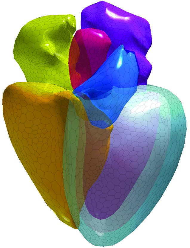
Philips Anatomical
Intelligence HeartModel tool
Intelligence HeartModel tool
With 23 hospitals and hundreds of health care facilities across southeast Wisconsin, the health system employs 19,600 individuals — some of whom have to travel to different facilities.
Through the implementation of the Centricity Cardio Enterprise, they have created a centralized view of cardiology images with customizable workflows and the ability to view related radiology images. It also allows for remote, web-based access to a patient’s imaging data across different specialties.
Ascension has experienced a dramatic decrease in turnaround time for cath lab reporting, according to Schowalter. The health system is conducting a scientific study to investigate this further and will present the results in May.
Schowalter and his team are evaluating the time it takes for the reports to get into the health systemwide electronic medical record (EMR) via dictation compared to using Centricity Cardio Enterprise.
“[CVIS] is almost a requirement now with all of the reporting that has to be done,” says Cris Bennett, a clinical analyst at MD Buyline. “All of the cath lab imaging data is stored in the CVIS, but it also reports stents and balloon pops and anything else they are going to use in a cath lab.”
Other CVIS solutions on the market include Philips CVIS and the Siemens Cardiovascular Imaging and Information Solution.
If a health care facility doesn’t have a dedicated CVIS, Bennett says it will embed the reporting into its PACS or any software in the radiology department that is compatible with HL7.
Promising research with micro-CT
Aarhus University in Denmark has developed a new contrast-enhanced micro-CT imaging technique that could one day produce 3-D reproductions of the cardiac conduction system, which generates the electrical signal that drives the heartbeat.
An article published in Scientific Reports in September stated that this technique allows for much higher spatial resolution than a conventional CT scanner. That will provide researchers with a better view of the cardiac conduction system and allow them to map the orientation of the cellular chains within the heart muscles that dictate the velocity and pathway of the electrical signal.
“Integrating these two pieces of structural information into mathematical simulations of the heart’s electrical activation can allow us to predict areas prone to arrhythmias and potentially find new targets for ablation therapy,” says Robert Stephenson, Marie Currie fellow at the university.
Knowing the 3-D disposition of the specialized cardiac conduction system can help surgeons with valve prosthesis implantation. It’s imperative that the prosthesis does not compress the neighboring specialized cells of the cardiac conduction system.
Currently, this technique is only performed on deceased patients for research purposes because the X-ray dose required to produce images of this quality is too high for clinical application. However, as CT technology improves, this could change.
Stephenson and his team have recently expanded the technique to explore the 3-D disposition of the cardiac conduction system of congenitally malformed human hearts. That information will help guide corrective surgery.
“We have also used the high resolution micro-CT data to produce 3-D prints of these hearts,” he says. “Such prints have potential implications in surgical planning and practice, medical education and even patient consultations.”
Making inroads into heart disease diagnosis
Two cardiac imaging technologies are in development that could open new avenues for predicting the risk of and diagnosing heart disease.
The EU’s Horizon 2020 collaboration is working on a handheld scanner that general practitioners can use during a routine health exam to spot patients with early-onset cardiovascular disease.
The device leverages a technique called Laser Doppler Vibrometry to create a vibration map of the chest and heart area. That map reveals signs of cardiovascular disease such as plaque build-up, arterial stiffness, arterial stenosis and heart dyssynchrony.
The current methods for diagnosing this condition are cardiac biomarkers, cardiac catheterization, chest X-ray, ECG, Holter monitoring and cardiac MR. Those approaches can be pricey, but the handheld scanner is expected to cost less than $2,000.
Medtronic has been appointed the technical coordinator of this collaboration and a prototype is expected to be unveiled this summer.
The University of Warwick in England, along with the Baker Heart and Diabetes Institute and Monash University in Australia, developed a laser imaging technique that can identify high-risk arterial plaques.
The team of researchers found that when they increased the wavelength of light that’s used to assess fatty build-up in arteries, they can identify rupture-prone deposits that often lead to blood clots, heart attack and stroke.
Current imaging techniques can identify some characteristics of high-risk plaques, but no method has been deemed reliable for selectively detecting these types of plaques. For instance, coronary angiography only detects narrow segments in the coronary artery.
If the laser imaging technique demonstrates positive results in clinical trials, it could one day be used to evaluate unstable fatty arterial plaques. It could also be used to monitor the effectiveness of drugs intended to prevent heart attack and stroke.