Duke-led study explores mouse brain anatomy with 64 million times greater resolution
Researchers at the Duke Center for In Vivo Microscopy have developed a new MR imaging method that increases resolution 64 million times over traditional MR scanners, giving scientists access to greater insights into Alzheimer's and other diseases by better observing changes in brain cells.
Using a vertical bore 9.4T Agilent (Varian) MR, gradient coils 100 times stronger than traditional ones, and a high-performance computer equivalent to the power of nearly 800 laptops, the researchers conducted their research on the brain of a mouse.
Collaborating with the researchers at Duke were scientists from the University of Tennessee Health Science Center, University of Pennsylvania, University of Pittsburgh and Indiana University, who compiled their findings in a paper, “Merged Magnetic Resonance and Light Sheet Microscopy of the Whole Mouse Brain.”
For those who need to move fast and expand clinical capabilities -- and would love new equipment -- the uCT 550 Advance offers a new fully configured 80-slice CT in up to 2 weeks with routine maintenance and parts and Software Upgrades for Life™ included.
They say that the record-breaking resolution enables them to visualize the wiring diagram of connections throughout the entire brain of a mouse, and could eventually allow them to study various drug interventions or dietary changes and the effects they have on cognitive awareness, as well as biological health, throughout aging and neurodegenerative disease processes.
"If we can give people even an extra year or two of cognitive awareness and health by looking at just modest interventions, the impact on the population and the cost of caring for aging patients would be enormous," lead author G. Allan Johnson, Charles E. Putman University Distinguished Professor of Radiology, Physics and Biomedical Engineering at Duke, told HCB News.
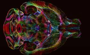
Once scanned, the mouse tissue was again imaged with light sheet microscopy, with both approaches allowing the researchers to label specific groups of cells across the brain, such as ones that produce dopamine and can be monitored with the new MR imaging solutions for signs of Parkinson’s disease.
They then mapped the light sheet pictures onto the original MR scans, creating more anatomically accurate images that provide vivid views of cells and circuits throughout the entire brain.
One set of scans showed how connectivity throughout the brain changed as the mice aged, as well as how specific regions, like the memory-involved subiculum, changed more than the rest of the brain. Another set showed rainbow-colored brain connections that indicated significant deterioration of neural networks in a mouse model for Alzheimer’s disease.