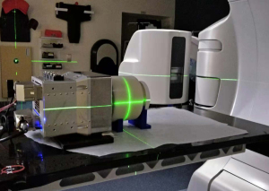
Michigan researchers have found a way to track and guide radiotherapy doses toward cancers more precisely using sound waves. (Photo courtesy of U-M Optical Imaging Laboratory)
In radiation therapy, X-rays heat up tissues when interacting with them, and create sound waves in the process. These tiny waves are weak and usually undetectable with ultrasound technology.
But researchers at the University of Michigan have found a way to use them to track radiotherapy treatments in real time with 3D imaging.
Using a new ionizing radiation acoustic imaging system (iRAI), they can capture and amplify sound waves to pinpoint radiation dose in the body, guiding clinicians to accurately direct radiation toward cancerous cells and away from adjacent, healthy tissue.
Such technology could be used to personalize each course of radiotherapy treatment, according to corresponding author Xueding Wang, the Jonathan Rubin Collegiate Professor of Biomedical Engineering, a radiology professor and head of the U-M’s Optical Imaging Laboratory at the University of Michigan, School of Medicine.
He told HCB News that the iRAI system maps dose deposited deep in the body by "listening" to sounds generated by the radiation beam without interfering with the treatment. “We have already started to test and validate this technique for clinical use. The initial experiments on patients have shown promising results. We plan to test the technique on more patients (>20), trying to further improve its performance and understand its clinical viability, advantages and limitations.”
The iRAI system uses an array of ultrasonic transducers positioned on the patient’s side to detect and amplify the sound waves. It then transfers the signal into an ultrasound device for image reconstruction, with providers using the scans to alter the level or trajectory of radiation.
The method is expected to be especially beneficial for when the target is adjacent to radiation-sensitive organs such as the small bowel or stomach. Additionally, the technology can be easily added on to current radiation therapy equipment without having to alter workflow processes. It can even be applied to improve accuracy and patient outcomes in advanced treatments like proton therapy, according to Wang.
“Proton therapy as well as other advanced radiation therapies can provide better accuracy than traditional X-ray radiation therapy. However, they are facing similar challenges such as the uncertainty in dose delivery due to the body motion or patient-to-patient difference,” he said.
The University of Michigan has applied for patent protection and is seeking partners to bring the technology to market.
The research was supported by the National Cancer Institute and the Michigan Institute for Clinical and Health Research.
Research on the technique was published in Nature Biotechnology.