by
John R. Fischer, Senior Reporter | June 17, 2022
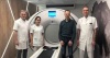
Using a Mid-IR beamline and infrared imaging can determine where and when hemorrhagic strokes begin. (Photo courtesy of Canadian Light Source)
A new approach may eventually enable MR and CT to show where bleeding for a hemorrhagic stroke starts and when it begins, helping clinicians make better care decisions for patients.
Such critical information can make a difference in patient outcomes, said researchers at the University of Saskatchewan and Curtin University in Australia. Using Fourier-transform infrared imaging, which produces an infrared spectrum that shows absorption or emission of solids, liquids or gasses, and a Mid-IR spectromicroscopy beamline from USask’s Canadian Light Source, they were able to observe oxidative damage in brain tissue samples associated with hemorrhagic stroke.
This is currently not feasible with a microscope or traditional imaging methods, according to Dr. Jake Pushie, a researcher at USask’s College of Medicine. "Conventional imaging generates a snapshot in time, with no way of knowing precisely when the bleed initiated if it is not hyperacute. Our methods still capture a snapshot, except the oxidatively damaged lipid markers may provide us with a 'clock', so to speak, at the site of injury that tells us how long ago the oxidative damage started to occur, which will be tied to the age of the hematoma," he told HCB News.


Ad Statistics
Times Displayed: 174592
Times Visited: 3185 For those who need to move fast and expand clinical capabilities -- and would love new equipment -- the uCT 550 Advance offers a new fully configured 80-slice CT in up to 2 weeks with routine maintenance and parts and Software Upgrades for Life™ included.
Knowing when bleeding stops can tell clinicians how much time they have to respond, because it helps them understand what is taking place biologically. This information could help doctors potentially minimize tissue damage faster.
Pushie and his colleagues plan to apply the technique on more stroke tissue samples to better determine the speed at which oxidative damage occurs. "Currently, we rely on anatomical snapshots in time and make clinical decisions based on generalized population-based concepts. This may not be the most applicable to the individual patient. Future imaging modalities, such as the work done by our team, can provide more patient-specific information on what is happening in their brains to help clinicians push the boundaries of time-limited therapies, such as surgery or medical management."
Several innovations are underway to improve detection of and treat strokes faster. Researchers at Mayo Clinic recently
found that using AI-enabled ECG for detecting atrial fibrillation can determine risk for cerebral strokes as well, due to their association with the former.
And engineers at the Massachusetts Institute of Technology have
developed a telerobotic system that allows surgeons to remotely control a robotic arm and safely operate on a patient during the “golden hour” of time to save their life and preserve brain function, even when the patient is in a completely separate location.
The findings of the Canadian and Australian study were published in
Metallomics.