From the November 2018 issue of HealthCare Business News magazine
On the back of the successful prototype, MARS Bioimaging Ltd. formed, with the goal of creating a commercial preclinical system, and scaling the technology for clinical use. “Modern imaging technologies – ultrasound, MR, CT and PET – were incrementally improving medical diagnosis year by year, but there were still many common diseases that couldn’t be diagnosed,” he said, “…and still cannot.”
Developing and marketing a preclinical system was a conscious decision to introduce a high-resolution color X-ray imaging system to researchers first, giving them a head start toward addressing current imaging challenges and answering the question of how this type of quantitative imaging can be used to go beyond current clinical imaging techniques.
The technology has come a long way in the past decade. “In the early days, a mouse took around 24 hours to scan and over a week to reconstruct,” said Butler. “Today, a mouse can be scanned in a matter of minutes, and reconstructed in an hour.”
The first challenge has always been to collect the best energy information possible. The second challenge is to best use that information to analyze the object. Then you need to produce images that convey that information to users.
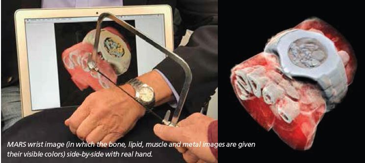
The energy information and the small pixel size enable MARS to produce lifelike images such as the image of Phil’s watch and half his hand, an image that went viral with tens of millions of reposts. Phil remarked, “I find the image beautiful, even though some find it horrid.”
“My turning ankle is less horrid perhaps, but contains more information, particularly if you seek out the high resolution images of the calcium, fatty, and water maps. We get exquisite separation of materials at the 80 micron, or 0.08 mm, scale.”
Looking to the future, MARS imaging has already demonstrated its usefulness for orthopedic joint scanning, and it is easy to see that within a few years, head and neck scanning will be viable. MARS scans of shoulders and hips will require methods to analyze and display terabyte data sets.
The preclinical research has demonstrated how transformative this technology will be in areas such as cancer detection, bone health, bone-metal interface imaging, and cardiovascular health assessment.