From the 19th century to present time, the X-ray has played a pivotal role in medical imaging. Similar to nearly any other technology, X-ray imaging has evolved significantly since its inception, most recently with the shift from analogue to digital.
For several years, analogue X-ray existed as the de facto standard in the market, but similar to other methods, its existence has been plagued with numerous problems and challenges. Not only has digital become the gold-standard in developed countries, but analogue's presence is slowly but surely disappearing in developing countries in place of new digital technology. In fact, the global digital X-ray market is estimated to be worth $4.82 billion by 2018, growing at a CAGR of 4.5% from 2013 to 2018, according to a recent Markets and Markets report.
Benefits of shifting from analogue to digital
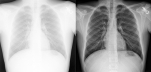
The ideal imaging system should permit a high quality image with minimal radiation exposure. Digital radiology has the potential to achieve this and further advances will possibly lead to lowering the radiation dose and using higher sensitivity plates to provide better resolution and sharpness of images.
The benefits of digital radiology are many; both financially, environmentally, and clinically. These include:
1. Providing high quality images, faster turn around and outcome, making it possible to review previous imaging on a patient much easier;
2. Minimal radiation exposure, for both patient and personnel;
3. Resource efficiency with less film cost and storage space and fewer staff handling the archive;
4. Opening up for future image processing and analysis improvements; and,
5. Sending and sharing difficult cases with colleagues can easily be done.
Minimizing radiation exposure
In the earlier days of X-ray when film-screen radiology was standard, the relation between blackness of the film and detector (film) exposure was clear. Every X-ray technologist could adjust the dose (mAs) to achieve the requested blackness of the film, so exposure was more precise and specific for each image.
Radiology has played an important role in the diagnosis and management of patients for more than 110 years. Traditional screen-film systems use overall film density as an exposure indicator. Direct feedback to the technologist regarding exposure is obtained by the appearance of the processed film image. Optimized technique factors (kVp and mAs) are based upon the patient size and body part and radiographic speed of the screen film combination being used. Particularly in situations where automatic exposure control is not used (for instance, in the majority of small pediatric patients), the use of fixed exposure parameters requires the technologist to use experience and appropriate judgment to set radiographic techniques.